Have you ever wondered about the intricate inner workings of the human body, far beyond what meets the eye? Human histology, the study of tissues at a microscopic level, holds the key to understanding the diverse structures that make up our organs and systems.
What is Human Histology?
Human histology delves deep into the microscopic world of cells and tissues that form the foundation of our body. By examining these tiny building blocks, researchers can uncover the secrets behind various physiological functions and pathological conditions.
The Fascinating World of Cells
Cells are the fundamental units of life, each with a specific role to play in maintaining the body’s equilibrium. From the powerhouse mitochondria to the protective skin cells, every cell contributes to the seamless functioning of our organs.
Types of Human Tissues
Human tissues are classified into four main types: epithelial, connective, muscle, and nervous tissue. Each type serves a unique purpose, whether it’s providing structural support, enabling movement, or transmitting electrical signals.
The Role of Histologists
Histologists are the unsung heroes behind the scenes, meticulously preparing tissue samples for examination under a microscope. Their keen eye for detail helps diagnose diseases and unravel the mysteries of cellular behavior.
Unveiling Disease Processes
Through histological analysis, medical professionals can identify abnormal tissue changes associated with diseases such as cancer, infections, and autoimmune disorders. This invaluable insight guides treatment decisions and improves patient outcomes.
Human histology may seem like a complex puzzle at first glance, but with the right tools and knowledge, we can unlock its secrets and gain a deeper appreciation for the marvels of the human body. So next time you marvel at the wonders of life, remember that the true magic lies beneath the surface, waiting to be discovered through the lens of histology.
About the Book
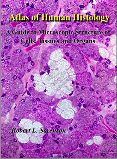
This atlas is a series of low-to-high magnification images of individual tissue specimens. The low-power images are used for orientation, while the higher-power images show cellular, tissue, and organ details. While every effort has been made to faithfully reproduce tissue coloration, a complete understanding of histological structure is best achieved by viewing the original specimen under a microscope.
This atlas provides a glimpse of what one should observe. It contains micrographs from a collection of microscopic slides that are used by medical, dental, and undergraduate students to study histology at the University of Minnesota. The majority of these slides were prepared by Anna-Mary Carpenter, M.D., Ph.D., during her time as a professor in the Department of Anatomy at the University of Minnesota Medical School.
Each tissue sample was digitized with a high-resolution 40x or 60x lens in its entirety, creating virtual microscope slides. The virtual microscope collection includes additional slides that complement and extend the core slide collection. The production of the Virtual Slide Collection and the development of the website for its presentation were done with the assistance of Todd C. Brelje, Ph.
The drawings in the Atlas are by Jean E. Magney, a gifted histologist and artist. Her gifted interpretation of biological structures and their artistic expression greatly enhances the study and understanding of histology. These drawings were first published in “Color Atlas of Histology, “Stanley L. Erlandsen and Jean E. Magney, Mosby 1992.

